Lipi Droplet Assay Kit (Blue, Deep Red)

Lipi Droplet Assay Kit (Blue, Deep Red)
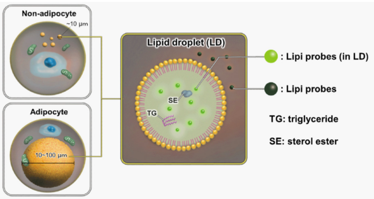
Lipiシリーズは脂肪親和性の高い低分子蛍光試薬であり、疎水性環境下で蛍光が増強します。試薬を添加するだけで生細胞および固定化細胞中の脂肪滴の量的変動を数値化することができます。
操作を大幅に短縮しており、生細胞にも使用可能。比色試薬を用いた方法と比較して、測定に必要な時間を大幅短縮、またプレートに色素が沈着しないため実験の再現性を高めることも可能。
製品情報
脂肪滴測定キット:数値化
Technical info
Lipid droplets (LDs) are composed of neutral lipids such as triacylglycerol & cholesteryl ester that are surrounded by phospholipid monolayers and are seen ubiquitously, not only in adipocytes1). Although LDs were simply thought to serve as a lipid storage unit, a recent study has stated that LDs play an important role in regulating lipid metabolism, autophagy2) and cellular senescence3).Therefore, have gained great attention as an important tool to elucidate the mechanisms of formation, growth, fusion, and retraction of LDs.

1) T. Fujimoto et al., “Lipid droplets: a classic organelle with new outfits.” Histochem Cell Biol., 2008, 130(2), 263.
2) R. Singh et al., “Autophagy regulates lipid metabolism.” Nature, 2009, 458(7242), 1131.
3) M. Yokoyama et al., “Inhibition of endothelial p53 improves metabolic abnormalities related to dietary obesity.” Cell Reports, 2014, 7(5), 1691.
Only by the addition of a reagent, the imaging of lipid droplets (LDs) or the quantitative variation of LDs in live and fixed cells becomes quantifiable.

Lipid Droplet Assay Kit considerably shortens the entire process and can be used for live cells.
The fluorescent dye provided in the Lipid Droplet Assay Kit can be used for live and fixed cells. Compared to a method of using a colorimetric reagent, the method of using the Lipid Droplet Assay Kit can shorten measuring time. Furthermore, the repeatability of experiment can be increased by using the Lipid Droplet Assay Kit because the dye is not deposited in a plate.

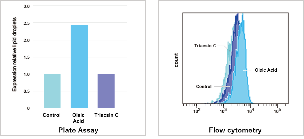
Experimental example of plate assay
Changes in lipid droplets were examined after the addition of oleic acid or Triacsin C (acyl-CoA synthetase inhibitor) to the A549 cell culture medium.
As a result, we confirmed that the oleic acid-treated cells show an increase in the number of LDs, compared to control and Triacsin C-treated cells.

Blue :Ex. 376 – 386 nm / Em 435 – 455 nm
Deep Red :Ex. 623 – 633 nm / Em 649 – 669 nm
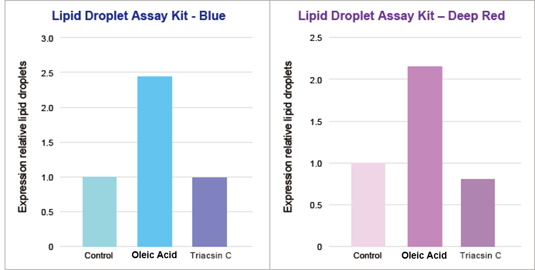
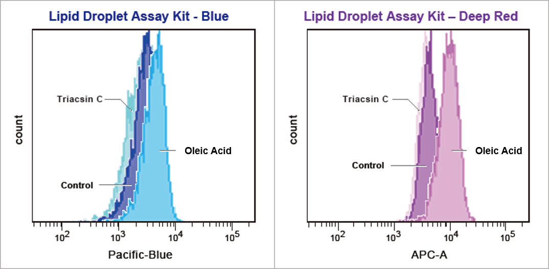
Reagent Comparison
Changes in lipid droplets were examined after the addition of oleic acid or Triacsin C (acyl-CoA synthetase inhibitor) to the HeLa cell culture medium.
As a result, we confirmed that the oleic acid-treated cells show an increase in the number of LDs, compared to control and Triacsin C-treated cells.

Blue :Ex. 405 nm/ Em 425 – 475 nm
Deep Red :Ex. 640 nm/ Em 650 – 670 nm
Related Product Information
Function: Imaging
Lipi-Blue 10 nmol LD01
Lipi-Green 10 nmol LD02
Lipi-Red 100 nmol LD03
Lipi-Deep Red 10 nmol LD04
Function: Quantification (Plate Reader, FCM)
Lipid Droplet Assay Kit - Blue 1 set LD05
Lipid Droplet Assay Kit - Deep Red 1 set LD06