GSSG/GSH Quantification Kit

GSSG/GSH Quantification Kit
酸化・還元型のグルタチオンを分けて測定。グルタチオン(γ-L-Glutamyl-L-cysteinylglycine)は生体内に存在するトリペプチドで、Glutathione peroxidase、glutathione S-transferaseおよびthiol transferase等の酵素基質として抗酸化や薬物代謝などに関与しています。グルタチオンは通常、生体内で還元型(GSH)として存在していますが、酸化ストレスなどの刺激によって還元型(GSH)から酸化型(GSSG)に変換されるため、GSHとGSSGの比率が酸化ストレスの指標として注目されています。
本製品は試薬を加えるだけの簡単操作で検査できます。
製品情報
グルタチオン定量キット
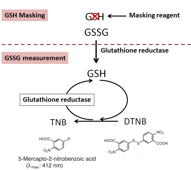
The GSSG/GSH Quantification kit contains Masking Reagent of GSH. The GSH can be deactivated in the sample by simply adding the Masking Reagent. Therefore, temp-only the GSSG is detected by measuring the absorption (λmax = 412 nm) of DTNB (5,5 Edithiobis (2-nitrobenzoic acid) using the enzymatic recycling system. Also, GSH can be determined the quantity by subtracting GSSG from the total amount of glutathione.
The kit is limited to quantifying GSH/GSSG concentration from 0.5 μmol/l to 50 μmol/l and GSSG concentration from 0.5 μmol/l to 25 μmol/l.
Developer Dojindo Molecular Technologies, Inc.
Technical info
Principle
Selective Quantification
Although conventional masking reagent, 2-Vinylpyridine(2-VP), interferes the reaction of GSSG measurement, Dojindo’s masking reagent does not interfere the reaction of GSSG measurement. Therefore, the exact ratio of GSSG and GSH is obtained with Dojindo GSSG/GSH detection Kit.

Measurement of GSSG with or without GSH masking reagent
Interference
Reducing agents such as ascorbic acid, β-mercaptoethanol, dithiothreitol (DTT) and cysteine, or thiol reactive compounds such as maleimide compounds, interfere with the glutathione assay. Therefore, SH compounds, reducing agents and SH reactive materials should be avoided during the sample preparation.
Required Equipment and Materials
– Plate reader (405 or 415 nm filter)
– 96-well microplate
– Incubator (37ºC)
– 20 µl and 200 µl pipettes, a multi channel pipette
– 5-Sulfosalicylic Acid (SSA) Solution
– Ethanol
Assay Procedure

Application
Literature used “GSSG/GSH Quantification Kit”
Diurnal Variation of cadmium-induced mortality in mice
N. Miura, Y. Yanagiba, K. Ohtani, M. Mita, M. Togawa, and T. Hasegawa, J. Toxicol. Sci., 2012, 37(1), 191
<Detailed information on this article>
– Measuring object: Hepatic GSH
– Sample: Liver (Mice)
– Preparation of sample
1) Homogenize liver sample in 5% SSA for 30 seconds in ice-water bath.
2) Centrifuge the homogenates consisting of 100 mg liver in 1 ml (10%) at 8,000 x g for 10 min. at 4°C to remove proteins.
3) Assay with supernatants for GSH using GSSG/GSH Quantification Kit according to the manufacture’s instruction.
Effect of Oxidative Stress on Secretory Function in Salivary Gland Cells
K. Okabayashi, T. Narita, Y. Takahashi, H. Sugiya, (2012), Oxidative Stress – Environmental Induction and Dietary Antioxidants, Edited by Volodymyr I. Lushchak, ISBN 978-953-51-0553-4, Hard cover, 388 pages, Publisher: InTech
<Detailed information on this article>
– Measuring object: GSH
– Sample: Parotid acinar cells
– Preparation of sample
1) Collect parotid acinar cells by centrifugation at 10,000 g for 15 s and immediately mixed with 160 ul of 10 mM HCl.
2) Freeze and thraw the mixture three times over, then mix with 40 ul of 5% SSA and then centrifuge at 8,000 g for 10 minutes.
3) Collect the supernatant and dilute twice.
Chloroplast DNA Replication Is Regulated by the Redox State Independently of Chloroplast Division in Chlamydomonas reinhardtii
Y. Kabeya, and S. Miyagishima, Plant Physiol., 2013, 161, 2102
<Detailed information on this article>
– Measuring object: GSH and GSSG
– Sample: Chlamydomonas reinhardtii
– Preparation of sample
1) Collect Chlamydomonas reinhardtii cells by 1,000g for 5 min and wash with PBS.
2) Resuspended in 5% SSA solution and disrupted by sonication.
3) Collect the supernatants by 15,000g for 5 min.
4) Isolate chloroplst by 700g for 5 min and wash with 50 mM HEPES-KOH, pH 7.5, containing 300mM sorbitol.
5) Resuspend in 5% SSA solution and centrifuge at 15,000g for 5 min
Selected publications
ER Stress Cooperates with Hypernutrition toTrigger TNF-Depe. Cancer Cell. 2014;26:331-343.
Purpose: Determine indicator of ER Stress in Liver
Mechanisms of cadmium-inducedchronotoxicity in mice. The journal of toxicological. 2013;38:947-957.
Purpose: Determine the GSH/GSSG ratio in cadmium exposed Liver
Hepatitis C Virus Core Protein SuppressesMitophagy by Interacting with Parkin in the Context of MitochondrialDepolarization. The American Journal of Pathology. 2014;184:3026-3039.
Purpose: Measure mitochondrial oxidative status in liver
Possible involvement of glutathione balance disruption in dihydropyrazine-induced cytotoxicity on human hepatoma HepG2 cells.The journal of toxicological.2012;37:1065-1069.
Purpose: Investigate fluctuation of GSH/GSSG ratio in the cytotoxicity HepG2 Cells
Induction of heme oxygenase-1 contributes to survival of Mycobacterium abscessus in humanmacrophages-likeTHP-1cells.Redox Biology. 2015;4:328-339.
Purpose: Determine redox state in macrophages
Age-related changes in salivary biomarkers.Journal of Dental Sciences. 2014;9:85-90.
Purpose: Measure GSH/GSSG ratio in salivary
Protective effects of hydrogen sulfideanions against acetaminophen-induced hepatotoxicity in mice. The journal oftoxicological. 2015;40:837-841.
Purpose: Measure glutathione of Hepatocyte
References
1. N. Kubota, et al., A high-fat diet and multiple administration of carbon tetrachloride induces liver injury and pathological features associated with non-alcoholic steatohepatitis in mice. Clin Exp Pharmacol Physiol. 2013; 40(7): 422-30
2. T. Tomofuji, et al., Supplementation of broccoli or Bifidobacterium longum-fermented broccoli suppresses serum lipid peroxidation and osteoclast differentiation on alveolar bone surface in rats fed a high-cholesterol diet. Nutr Res. 2012; 32(4): 301-7
3. T. Miyayama., et al., Mitochondrial electron transport is inhibited by disappearance of metallothionein in human bronchial epithelial cells following exposure to silver nitrate. Toxicology. 2013; 8;305: 20-9.
4. H. Miwa, et al., Leukemia cells demonstrate a differen t metabolic perturbation provoked by 2-deoxyglucose. Oncol Rep. 2013; 29(5): 2053-7.
5. K. Unno, et al., Acute enhancement of non-rapid eye movement sleep in rats after drinking water contaminated with cadmium chloride. J Appl Toxicol. 2013 24.
6. Y. Ishihara, et al., Tributyltin induces oxidative stress and neuronal injury by inhibiting glutathione S-transferase in rat organotypic hippocampal slice cultures. Neurochem Int. 2012; 60(8): 782-90.
7. H. Miyamoto, et al., Thermophile-fermented compost as a possible scavenging feed additive to prevent peroxidation. J Biosci Bioeng. 2013; 116(2): 203-8.
8. E. Taniai, et al., Ochratoxin A induces karyomegaly and cell cycle aberrations in renal tubular cells without relation to induction of oxidative stress responses in rats. Toxicology Letters, October 2013.
9. G. Tian, et al., Ubiquinol-10 Supplementation Activates Mitochondria Functions to Decelerate Senescence in Senescence Accelerated Mice. Antioxidants & Redox Signaling, October 2013.
Data

参考文献
2) M. A. Baker, G. J. Cerniglia and A. Zaman, "Microtiter Plate Assay for the Measurement of Glutathione and Glutathione Disulfide in Large Numbers of Biological Samples", Anal. Biochem., 1990, 190, 360.
3) C. Vandeputte, I. Guizon, I. Genestie-Denis, B. Vannier and G. Lorenzon, "A Micrototer Plate Assay for Total Glutathione and Glutathione Disulfide Contents in Cultured/isolated Cells: Performance Study of a New Miniaturized Protocol", Cell Biol. Toxicol., 1994, 10, 415.
4) S. A. McGrath-Morrow and J. Stahl, "Inhibition of Glutamine Synthetase in A549 Cells During Hyperoxia", Am. J. Respir. Cell Mol. Biol., 2002, 27, 99.
5) T. Sato, K. Seyama, Y. Sato, H. Mori, S. Souma, T. Akiyoshi, Y. Kodama, T. Mori, S. Goto, K. Takahashi, Y. Fukuchi, N. Maruyama and A. Ishigami, "Senescence Marker Protein-30 Protects Mice Lungs from Oxidative Stress, Aging, and Smoking", Am. J. Respir. Crit. Care Med., 2006, 174, 530.
6) M. L. Mulhern, C. J. Madson, A. Danford, K. Ikesugi, P. F. Kador and T. Shinohara, "The Unfolded Protein Response in Lens Epithelial Cells from Galactosemic Rat Lenses", Invest. Ophthalmol. Vis. Sci., 2006, 47(9), 3951.
7) N. Miura, Y. Yanagiba, K. Ohtani, M. Mita, and M. Togawa, "Diurnal Variation of Cadmium-induced Mortality in Mice", J. Toxicol. Sci., 2012, 37(1), 191.
8) K. Okabayashi, T. Narita, Y. Takahashi and H. Sugiya, "Effrct of Oxidative Stress on Secretory Function in Salivary Gland Cells", Oxidative Stress-Environmental Induction and Dietary Antioxidants, V. Lushchak, InTech, 2012, 189.
9) G. Tian, J. Sawashita, H. Kubo, S. Nishio, S. Hashimoto, N. Suzuki, H. Yoshimura, M. Tsuruoka, Y. Wang, Y. Liu, H. Luo, Z. Xu, M. Mori, M. Kitano, K. Hosoe, T. Takeda, S. Usami and K. Higuchi, "Ubiquinol-10 Supplementation Activates Mitochondria Functions to Decelerate Senescence in Senescence-Accelerated Mice", Antioxid. Redox Signal., 2014, 20(16), 2606.
10) H. Nakagawa, A. Umemura, K. Taniguchi, J. Font-Burgada, D. Dhar, H. Ogata, Z. Zhong, M. A. Valasek, E. Seki, J. Hidalgo, K. Koike, R. J. Kaufman and M. Karin, "ER Stress Cooperates with Hypernutrition to Trigger TNF-Dependent Spontaneous HCC Development", Cancer cell, 2014, 26(3), 331.
11) M. Sueyoshi, M. Fukunaga, M. Mei, A. Nakajima, G. Tanaka, T. Murase, Y. Narita, S. Hirata, and D. Kadowaki, "Effects of lactulose on renal function and gut microbiota in adenine-induced chronic kidney disease rats", Clinical and Experimental Nephrology., 2019,doi: 10.1007/s10157-019-01727-4.
12) S. Shiromizu, T. Yamauchi, N. Kusunose, N. Matsunaga, S. Koyanagi and S. Ohdo, Dosing Time-Dependent Changes in the Anti-tumor Effect of xCT Inhibitor Erastin in Human Breast Cancer Xenograft Mice', Biol. Pharm. Bull.., 2019, 42, (11), 1921-1925.
%20--%3e%3csvg%20version='1.1'%20id='レイヤー_1'%20xmlns='http://www.w3.org/2000/svg'%20xmlns:xlink='http://www.w3.org/1999/xlink'%20x='0px'%20y='0px'%20viewBox='0%200%2055%2063.3'%20style='enable-background:new%200%200%2055%2063.3;'%20xml:space='preserve'%3e%3cstyle%20type='text/css'%3e%20.st0{fill:none;}%20.st1{fill:none;stroke:%23FFFFFF;stroke-miterlimit:10;}%20.st2{fill:%23EAADC6;}%20%3c/style%3e%3cg%20id='レイヤー_2_00000074435297100839857730000016701895290514446243_'%3e%3cg%20id='レイヤー_1-2'%3e%3crect%20class='st0'%20width='55'%20height='63.3'/%3e%3cg%20id='Layer_1'%3e%3cg%3e%3cpath%20class='st1'%20d='M16.3,6.3h22.5c1.6,0,3,1.3,3,3v50.1c0,1.6-1.3,3-3,3H16.3c-1.6,0-3-1.3-3-3V9.2%20C13.3,7.6,14.6,6.3,16.3,6.3z'/%3e%3cpath%20class='st1'%20d='M21.9,1h11.3c0.6,0,1.1,0.5,1.1,1.1v4.2H20.8V2.1C20.8,1.5,21.3,1,21.9,1z'/%3e%3crect%20x='16.2'%20y='9.9'%20class='st2'%20width='22.2'%20height='7.7'/%3e%3crect%20x='16.2'%20y='51.7'%20class='st2'%20width='22.2'%20height='7.7'/%3e%3cpolygon%20class='st2'%20points='29.9,19.9%2022.5,34.5%2026.9,36.4%2024.8,48.8%2032.5,33.9%2027.8,32.1%20'/%3e%3cg%3e%3cline%20class='st1'%20x1='25.2'%20y1='13.8'%20x2='29.4'%20y2='13.8'/%3e%3cline%20class='st1'%20x1='27.4'%20y1='11.7'%20x2='27.4'%20y2='15.9'/%3e%3c/g%3e%3cline%20class='st1'%20x1='25.2'%20y1='55.5'%20x2='29.4'%20y2='55.5'/%3e%3c/g%3e%3c/g%3e%3c/g%3e%3c/g%3e%3c/svg%3e)
%20rotate(-58.77)'/%3e%3cellipse%20class='cls-2'%20cx='28.07'%20cy='31.88'%20rx='31.38'%20ry='12.11'%20transform='translate(-12.46%2019.17)%20rotate(-31.23)'/%3e%3ccircle%20class='cls-1'%20cx='19.93'%20cy='9.05'%20r='3.48'/%3e%3ccircle%20class='cls-1'%20cx='51.74'%20cy='38.02'%20r='3.48'/%3e%3ccircle%20class='cls-1'%20cx='16.88'%20cy='48.55'%20r='3.48'/%3e%3ccircle%20class='cls-1'%20cx='28.07'%20cy='31.88'%20r='5.14'/%3e%3c/g%3e%3c/g%3e%3c/svg%3e)
