Glutamate Assay Kit-WST
Glutamate Assay Kit-WST
細胞内の代謝システムである、回答系やTCA回路、ペントース-リン酸経路の解析は、細胞状態を理解するうえで重要であり、グルコースや乳酸、NAD(P)*/NAD(P)Hなどのエネルギーおよび代謝産物を指標に評価されています。
製品情報
細胞内代謝測定(グルタミン酸)
Our Glutamate Assay Kit-WST is designed to quantify glutamate as a metabolite. This kit allows you to quantify glutamate present in culture medium or intracellular glutamate by WST reduction reaction. The lowest concentration of glutamate which can be quantified is 5 μmol/l. This kit can be used with 96-well microplates, which makes it possible to analyze multiple specimens.
Technical info
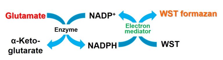
The kit is used to detect Glutamate in cell culture medium by measuring the absorbance of WST formazan produced according to quantities of glutamate. The kit includes a “Glutamate Standard” which can be used to quantify the concentration of lactate found within samples by creating standard curve.
Simple Assay Procedure
Procedures are so easy that you simply incubate the plate after the addition of culture supernatant or tissue/cell lysates prior to adding a reagent.

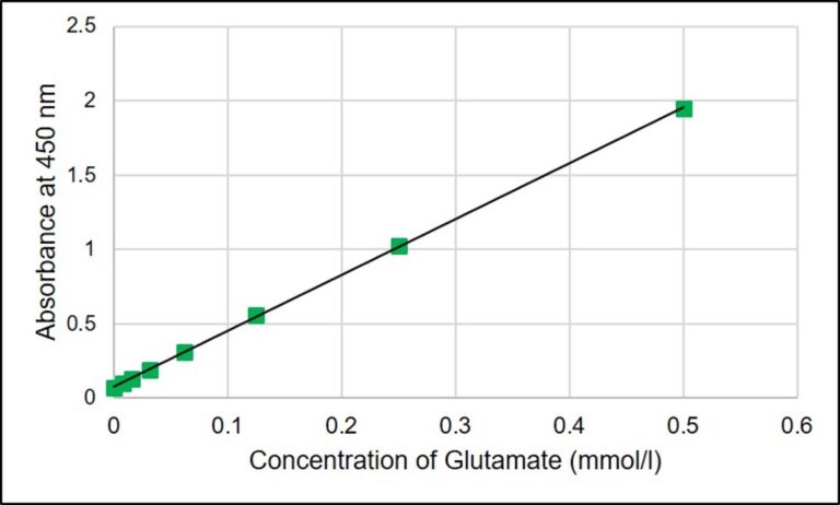
Standard Curve
Standard curve can be prepared by using Glutamate Standard included in the kit. The concentration of Glutamate can be measured. The concentration of lactate can be evaluated by diluting the samples if the concentration is over 0.5 mmol/l.
Example of Measurements of Glutamine/Glutamate level
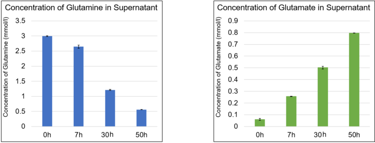

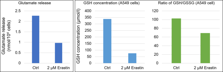
Example of Measurements of Glutamine/Glutamate level in Ferroptosis research
It is known that an iron-dependent cell death called “ferroptosis” is induced when xCT is inhibited by elastin treatment. The amounts of glutamate release and intracellular glutathione were measured by using the elastin-treated A549 cells. As a result, the amount of glutamate release was decreased in the elastin-treated A549 cells, and the amount of intracellular glutathione was decreased by inhibiting cystine uptake.

%20--%3e%3csvg%20version='1.1'%20id='レイヤー_1'%20xmlns='http://www.w3.org/2000/svg'%20xmlns:xlink='http://www.w3.org/1999/xlink'%20x='0px'%20y='0px'%20viewBox='0%200%2055%2063.3'%20style='enable-background:new%200%200%2055%2063.3;'%20xml:space='preserve'%3e%3cstyle%20type='text/css'%3e%20.st0{fill:none;}%20.st1{fill:none;stroke:%23FFFFFF;stroke-miterlimit:10;}%20.st2{fill:%23EAADC6;}%20%3c/style%3e%3cg%20id='レイヤー_2_00000074435297100839857730000016701895290514446243_'%3e%3cg%20id='レイヤー_1-2'%3e%3crect%20class='st0'%20width='55'%20height='63.3'/%3e%3cg%20id='Layer_1'%3e%3cg%3e%3cpath%20class='st1'%20d='M16.3,6.3h22.5c1.6,0,3,1.3,3,3v50.1c0,1.6-1.3,3-3,3H16.3c-1.6,0-3-1.3-3-3V9.2%20C13.3,7.6,14.6,6.3,16.3,6.3z'/%3e%3cpath%20class='st1'%20d='M21.9,1h11.3c0.6,0,1.1,0.5,1.1,1.1v4.2H20.8V2.1C20.8,1.5,21.3,1,21.9,1z'/%3e%3crect%20x='16.2'%20y='9.9'%20class='st2'%20width='22.2'%20height='7.7'/%3e%3crect%20x='16.2'%20y='51.7'%20class='st2'%20width='22.2'%20height='7.7'/%3e%3cpolygon%20class='st2'%20points='29.9,19.9%2022.5,34.5%2026.9,36.4%2024.8,48.8%2032.5,33.9%2027.8,32.1%20'/%3e%3cg%3e%3cline%20class='st1'%20x1='25.2'%20y1='13.8'%20x2='29.4'%20y2='13.8'/%3e%3cline%20class='st1'%20x1='27.4'%20y1='11.7'%20x2='27.4'%20y2='15.9'/%3e%3c/g%3e%3cline%20class='st1'%20x1='25.2'%20y1='55.5'%20x2='29.4'%20y2='55.5'/%3e%3c/g%3e%3c/g%3e%3c/g%3e%3c/g%3e%3c/svg%3e)
%20rotate(-58.77)'/%3e%3cellipse%20class='cls-2'%20cx='28.07'%20cy='31.88'%20rx='31.38'%20ry='12.11'%20transform='translate(-12.46%2019.17)%20rotate(-31.23)'/%3e%3ccircle%20class='cls-1'%20cx='19.93'%20cy='9.05'%20r='3.48'/%3e%3ccircle%20class='cls-1'%20cx='51.74'%20cy='38.02'%20r='3.48'/%3e%3ccircle%20class='cls-1'%20cx='16.88'%20cy='48.55'%20r='3.48'/%3e%3ccircle%20class='cls-1'%20cx='28.07'%20cy='31.88'%20r='5.14'/%3e%3c/g%3e%3c/g%3e%3c/svg%3e)
