Glucose Assay Kit-WST
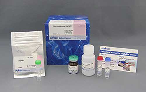
Glucose Assay Kit-WST
細胞内の代謝システムである、回答系やTCA回路、ペントース-リン酸経路の解析は、細胞状態を理解するうえで重要であり、グルコースや乳酸、NAD(P)*/NAD(P)Hなどのエネルギーおよび代謝産物を指標に評価されています。
製品情報
細胞内代謝測定(グルコース)
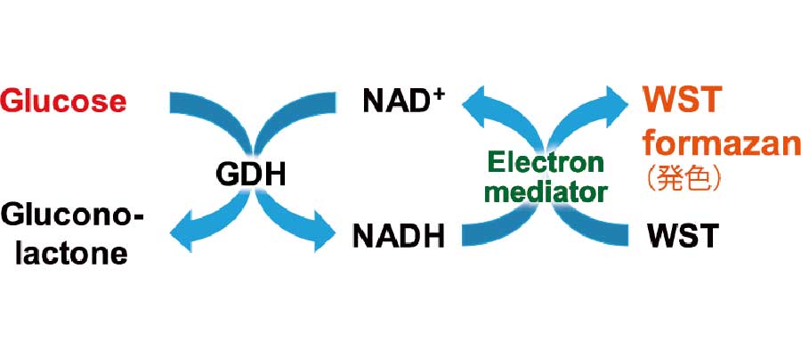
This kit can detect glucose found within cell culture medium or intracellular glucose by measuring the absorbance of the colored WST formazan dye. The degree of absorbance depends on the amount of glucose present within the sample.This kit contains a glucose standard solution that can be used to create a standard curve. This allows for the quantitation of glucose levels present in the sample.
Technical info
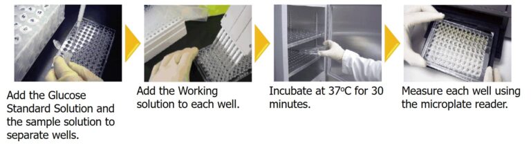
Procedure
Procedures are so easy that you simply incubate the plate after the addition of culture supernatant or tissue/cell lysates prior to adding a reagent.
Preparation of Standard Curve
Glucose levels in a sample can be measured by a calibration curve established with Glucose Standards included in this kit. If the glucose levels are greater than or equal to 0.5 mmol/L, the sample must be diluted before measurement.

Measurement of Glucose level in combination with Lactate Assay Kit
By using Glucose Assay Kit-WST and Lactate Assay Kit-WST, we have successfully measured metabolic activity changes of phloretin, a protein transport inhibitor, when it is added to Jurkat cells.

Precautions when using this kit.
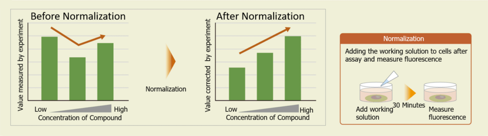
Cell counts may need to be normalized. When cells are analyzed in a microplate, the results obtained may sometimes differ depending on cell numbers per well.In such cases, normalization of the measured values obtained from cell counting and total protein will be necessary.In this kit, cell numbers can be easily measured by the fluorescence intensity induced by a reagent added to cell culture medium for staining nuclei.
参考文献
Cite: M. Shinohara, Y. Tashiro, M. Shinohara, J. Hirokawa, K. Suzuki, M. O. Takeya, M. Mukouzono, S. Takeda, T. Saito, A. Fukumori, T. C. Saido, R. Morishita and N. Sato, "Increased levels of Aβ42 decrease the lifespan of ob/ob mice with dysregulation of microglia and astrocytes”, FASEB J., 2019,DOI: 10.1096/fj.201901028RR
2) SampleMicroorganism(Streptomyces albulus )
Cite: K. Yamanaka, Y. Hamano and T. Oikawa, "Enhancement of metabolic flux toward ε-poly-l-lysine biosynthesis by targeted inactivation of concomitant polyene macrolide biosynthesis in Streptomyces albulus.”, J. Biosci. Bioeng., 2020,DOI: 10.1016/j.jbiosc.2019.12.002
3) Sample: Cell(HCT116)
Cite: K. Ohshima, S. Nojima, S. Tahara, M. Kurashige, K. Kawasaki, Y. Hori, M. Taniguchi, Y. Umakoshi, D. Okuzaki, N. Wada, J. Ikeda, E. Fukusaki and E. Morii, "Serine racemase enhances growth of colorectal cancer by producing pyruvate from serine”, Nat Metab, 2020, 2(1), 81
4) Sample: Cell(P388 Leukemia)
Cite: T. Matsuo, Y. Konya, E. Hirayama and Y. Sadzuka , "2-Deoxy-D-glucose enhances the anti-cancer effects of idarubicin on idarubicin-resistant P388 leukemia cells”, Oncol Lett , 2020, 20(1), 962-966
5) Sample: Cells (mouse: sperm)
Cite: M. Hashimoto, S. Kimura, C. Kanno, Y. Yanagawa, T. Watanabe, J. Okabe, E. Takahashi, M. Nagano and H. Kitamura, "Macrophage ubiquitin‑specific protease 2 contributes to motility, hyperactivation, capacitation, and in vitro fertilization activity of mouse sperm”, Cellular and Molecular Life Sciences, 2020, doi: 10.1007/s00018-020-03683-9
6) Sample: Cells. (Macrophages)
Cite: N. Saeki and Y. Imai, "Reprogramming of synovial macrophage metabolism by synovial fbroblasts under infammatory conditions ”, Cell Commun Signal, 2020, 18, 188
%20--%3e%3csvg%20version='1.1'%20id='レイヤー_1'%20xmlns='http://www.w3.org/2000/svg'%20xmlns:xlink='http://www.w3.org/1999/xlink'%20x='0px'%20y='0px'%20viewBox='0%200%2055%2063.3'%20style='enable-background:new%200%200%2055%2063.3;'%20xml:space='preserve'%3e%3cstyle%20type='text/css'%3e%20.st0{fill:none;}%20.st1{fill:none;stroke:%23FFFFFF;stroke-miterlimit:10;}%20.st2{fill:%23EAADC6;}%20%3c/style%3e%3cg%20id='レイヤー_2_00000074435297100839857730000016701895290514446243_'%3e%3cg%20id='レイヤー_1-2'%3e%3crect%20class='st0'%20width='55'%20height='63.3'/%3e%3cg%20id='Layer_1'%3e%3cg%3e%3cpath%20class='st1'%20d='M16.3,6.3h22.5c1.6,0,3,1.3,3,3v50.1c0,1.6-1.3,3-3,3H16.3c-1.6,0-3-1.3-3-3V9.2%20C13.3,7.6,14.6,6.3,16.3,6.3z'/%3e%3cpath%20class='st1'%20d='M21.9,1h11.3c0.6,0,1.1,0.5,1.1,1.1v4.2H20.8V2.1C20.8,1.5,21.3,1,21.9,1z'/%3e%3crect%20x='16.2'%20y='9.9'%20class='st2'%20width='22.2'%20height='7.7'/%3e%3crect%20x='16.2'%20y='51.7'%20class='st2'%20width='22.2'%20height='7.7'/%3e%3cpolygon%20class='st2'%20points='29.9,19.9%2022.5,34.5%2026.9,36.4%2024.8,48.8%2032.5,33.9%2027.8,32.1%20'/%3e%3cg%3e%3cline%20class='st1'%20x1='25.2'%20y1='13.8'%20x2='29.4'%20y2='13.8'/%3e%3cline%20class='st1'%20x1='27.4'%20y1='11.7'%20x2='27.4'%20y2='15.9'/%3e%3c/g%3e%3cline%20class='st1'%20x1='25.2'%20y1='55.5'%20x2='29.4'%20y2='55.5'/%3e%3c/g%3e%3c/g%3e%3c/g%3e%3c/g%3e%3c/svg%3e)
%20rotate(-58.77)'/%3e%3cellipse%20class='cls-2'%20cx='28.07'%20cy='31.88'%20rx='31.38'%20ry='12.11'%20transform='translate(-12.46%2019.17)%20rotate(-31.23)'/%3e%3ccircle%20class='cls-1'%20cx='19.93'%20cy='9.05'%20r='3.48'/%3e%3ccircle%20class='cls-1'%20cx='51.74'%20cy='38.02'%20r='3.48'/%3e%3ccircle%20class='cls-1'%20cx='16.88'%20cy='48.55'%20r='3.48'/%3e%3ccircle%20class='cls-1'%20cx='28.07'%20cy='31.88'%20r='5.14'/%3e%3c/g%3e%3c/g%3e%3c/svg%3e)
