Cellular Senescence Plate Assay Kit - SPiDER-βGal
Cellular Senescence Plate Assay Kit - SPiDER-βGal
細胞の生存および死をコントロールするために備わった機能として、アポトーシスやネクローシス、オートファジーは、細胞内機能を理解する上で非常に重要です。がん化因子として知られるSASPの発見や、Stem cell分野での老化現象の発見が認められるなど、各分野で重要視されています。細胞老化を評価するときには、複数の老化細胞マーカーを指標に解析する必要があります。
製品情報
老化細胞検出試薬・キット(プレートリーダーによる簡便な検出)
Technical info
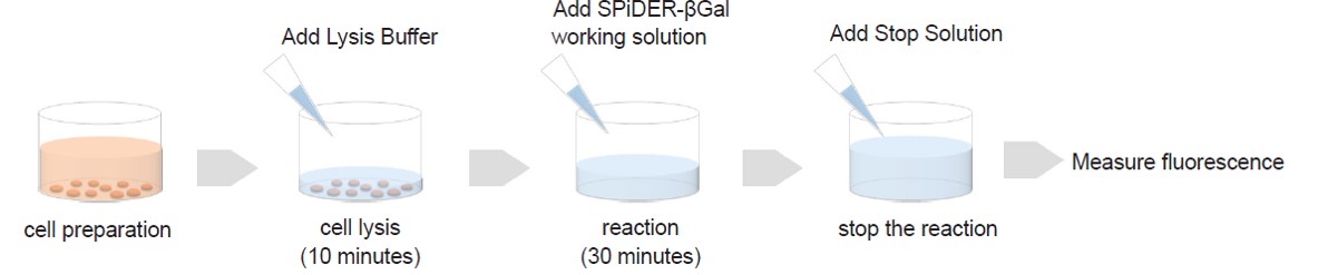
Cells prepared in advance are lysed in the buffer supplied with this kit.Fluorescence intensity is obtainable according to SA-β-gal activity simply by adding the fluorescent substrate SPiDER-βGal to the cell lysate. Even when you prepare cells in 100 mm dishes or others, fluorescence intensity can be measured by transferring cell lysate in 96 well plates after cell lysis.
Correlation with imaging data:
Imaging assessments of WI-38 cells at different passage levels were performed with this Plate Assay Kit and the Cellular Senescence Detection Kit – SPiDER-βGal
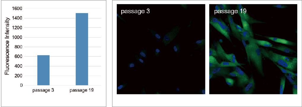
As a result, it was confirmed that in both kits, SA-β-gal staining increased in the high-passage WI-38 cells.
Bear in mind that although initial cell seeding densities are the same, cell densities at the time of plate assay differ due to low proliferation rate of senescent cells at higher passage levels. Therefore, in this experiment, we used SA-β-Gal activity values normalized by the results obtained using the Cell Count Normalization Kit (coming soon) in which cell number is determined by a nuclear marker.
Plate Assay
Ex. 535nm / Em. 580nm
Imaging data
Green: Ex. 488nm / Em. 500-600nm (SA-β-Gal staining with Cellular Senescence Detection Kit – SPiDER-βGal(Item# SG04))
Blue: Ex. 405nm / Em. 450-495nm (Nuclear staining with -Cellstain- DAPI solution(Item# D523))
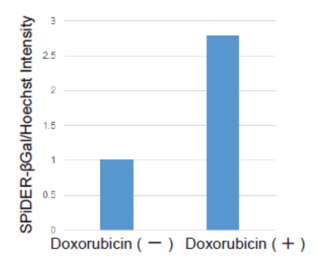
Evaluation doxorubicin-treated with cells
Cell counts may need to be normalized. When cells are analyzed in a microplate, the results obtained may sometimes differ depending on cell numbers per well.In such cases, normalization of the measured values obtained from cell counting and total protein will be necessary. In this kit, cell numbers can be easily measured by the fluorescence intensity induced by a reagent added to cell culture medium for staining nuclei.
Ex. 535nm / Em. 580nm
Precautions when using this kit
Cell counts may need to be normalized. When cells are analyzed in a microplate, the results obtained may sometimes differ depending on cell numbers per well.In such cases, normalization of the measured values obtained from cell counting and total protein will be necessary. In this kit, cell numbers can be easily measured by the fluorescence intensity induced by a reagent added to cell culture medium for staining nuclei.

%20--%3e%3csvg%20version='1.1'%20id='レイヤー_1'%20xmlns='http://www.w3.org/2000/svg'%20xmlns:xlink='http://www.w3.org/1999/xlink'%20x='0px'%20y='0px'%20viewBox='0%200%2055%2063.3'%20style='enable-background:new%200%200%2055%2063.3;'%20xml:space='preserve'%3e%3cstyle%20type='text/css'%3e%20.st0{fill:none;}%20.st1{fill:none;stroke:%23FFFFFF;stroke-miterlimit:10;}%20.st2{fill:%23EAADC6;}%20%3c/style%3e%3cg%20id='レイヤー_2_00000074435297100839857730000016701895290514446243_'%3e%3cg%20id='レイヤー_1-2'%3e%3crect%20class='st0'%20width='55'%20height='63.3'/%3e%3cg%20id='Layer_1'%3e%3cg%3e%3cpath%20class='st1'%20d='M16.3,6.3h22.5c1.6,0,3,1.3,3,3v50.1c0,1.6-1.3,3-3,3H16.3c-1.6,0-3-1.3-3-3V9.2%20C13.3,7.6,14.6,6.3,16.3,6.3z'/%3e%3cpath%20class='st1'%20d='M21.9,1h11.3c0.6,0,1.1,0.5,1.1,1.1v4.2H20.8V2.1C20.8,1.5,21.3,1,21.9,1z'/%3e%3crect%20x='16.2'%20y='9.9'%20class='st2'%20width='22.2'%20height='7.7'/%3e%3crect%20x='16.2'%20y='51.7'%20class='st2'%20width='22.2'%20height='7.7'/%3e%3cpolygon%20class='st2'%20points='29.9,19.9%2022.5,34.5%2026.9,36.4%2024.8,48.8%2032.5,33.9%2027.8,32.1%20'/%3e%3cg%3e%3cline%20class='st1'%20x1='25.2'%20y1='13.8'%20x2='29.4'%20y2='13.8'/%3e%3cline%20class='st1'%20x1='27.4'%20y1='11.7'%20x2='27.4'%20y2='15.9'/%3e%3c/g%3e%3cline%20class='st1'%20x1='25.2'%20y1='55.5'%20x2='29.4'%20y2='55.5'/%3e%3c/g%3e%3c/g%3e%3c/g%3e%3c/g%3e%3c/svg%3e)
%20rotate(-58.77)'/%3e%3cellipse%20class='cls-2'%20cx='28.07'%20cy='31.88'%20rx='31.38'%20ry='12.11'%20transform='translate(-12.46%2019.17)%20rotate(-31.23)'/%3e%3ccircle%20class='cls-1'%20cx='19.93'%20cy='9.05'%20r='3.48'/%3e%3ccircle%20class='cls-1'%20cx='51.74'%20cy='38.02'%20r='3.48'/%3e%3ccircle%20class='cls-1'%20cx='16.88'%20cy='48.55'%20r='3.48'/%3e%3ccircle%20class='cls-1'%20cx='28.07'%20cy='31.88'%20r='5.14'/%3e%3c/g%3e%3c/g%3e%3c/svg%3e)
