NADP/NADPH Assay Kit-WST

NADP/NADPH Assay Kit-WST
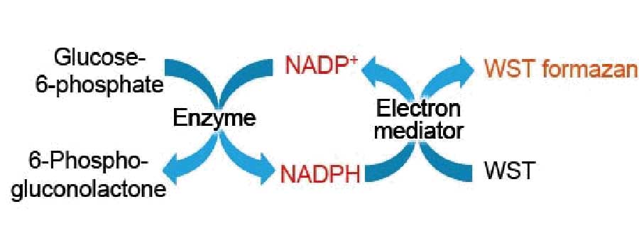
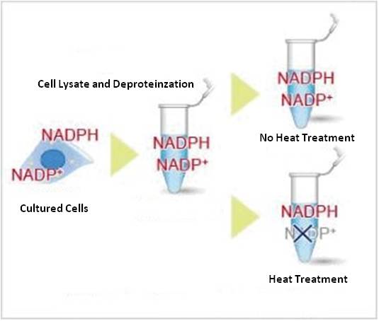
Cell lysate from a cell culture can be easily prepared via Deproteinzation using the extraction buffer and filtration tubes found within this kit. Intracellular NADPH levels can be quantified by heat treatment of cell lysate. Additionally, intracellular NADP+ levels can be determined by subtracting NADPH levels from independently measured total NADP+/NADPH levels.
NADP/NADPH Assay Kit-WST enables quantitation of the amount of total NADP+/NADPH, NADPH and NADP+ in cells and measurement of their ratio.
Product information
Intracellular metabolism measurement (NAD/NADH)
NADP/NADPH Assay Kit-WST enables quantitation of the amount of total NADP+/NADPH, NADPH and NADP+ in cells and measurement of their ratio.

Technical info

Cell lysate from a cell culture can be easily prepared via Deproteinzation using the extraction buffer and filtration tubes found within this kit. Intracellular NADPH levels can be quantified by heat treatment of cell lysate. Additionally, intracellular NADP+ levels can be determined by subtracting NADPH levels from independently measured total NADP+/NADPH levels.
This kit can be used to measure up to 12 samples and 8 standard samples (n=3). It is necessary to prepare additional filtration tubes when measuring more than 12 samples.
Study of NADP+/NADPH as Markers

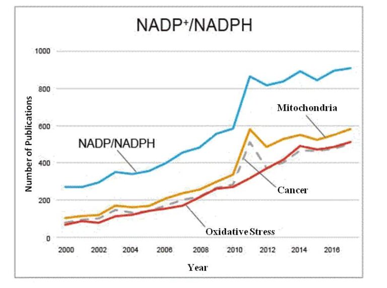
The intracellular levels of NADP+ and NADPH are recognized as important metabolic markers in understanding how cancer cells & mitochondrial function are influenced by drug administration, genetic engineering, etc. In particular, the number of publications has increased, mainly within the United States, indicating that the assessment of intracellular metabolism is considered important to understanding cellular states.
Producing Accurate Data in Plate Assay
The quantitative analysis of NADPH and total NADP+/NADPH levels (0.01-1.00 μmol/l) is possible with the simultaneous measurement of the standard buffer supplied with this kit. When the total NADP+/NADPH levels in samples are higher than 1 μmol/l, the quantitative analysis becomes possible with the use of the diluted samples.
It has been confirmed that the NADP/NADPH Assay Kit-WST reacts with neither NAD+ nor NADH (see right diagram below).

Example of Measurements in Combination with Glutathione (GSH) Quantification Kit
Change in metabolic activity was examined when anticancer drug doxorubicin (Dox) was added to Jurkat cells.
Dox was added to Jurkat cells (3×10⁶ cells) to obtain a final concentration of 500 nmol/l Dox, and after 24 hours incubation, NADP+/NADPH ratio and reduced/oxidized glutathione (GSH/GSSG) ratio were determined using the NADP/NADPH Assay Kit-WST and the GSSG/GSH Quantification Kit (Item#: G257), respectively, after the cells were washed in phosphate buffered saline (PBS).

The results shown above are likely to be explained by the following mechanism. When DOX (doxorubicin) was added to cells, DOX radicals, along with NADP+, were generated by enzymatic reaction. DOX radicals form reactive oxygen species (ROS), which induces DNA damage and apoptosis.In the meantime, to eliminate ROS formed in cells, GSH is consumed and GSSG is increased.Moreover, NADPH is used to reduce GSSG to GSH, resulting in an increase of NADP+.
References
2) S. Mandziuk, R. Gieroba, A. Korga, W. Matysiak, B. Jodlowska-Jedrych, F. Burdan, E. Poleszak, M. Kowalczyk, L. Grzycka-Kowalczyk, E. Korobowicz, A. Jozefczyk and J. Dudka, “The differential effects of green tea on dose-dependent doxorubicin toxicity.”, Food Nutr Res., 2015, 59, 29754.
3) M. Nagaya, H. Hara, T. Kamiya and T. Adachi,"Inhibition of NAMPT markedly enhances plasma-activated medium-induced cell death in human breast cancer MDA-MB-231 cells.", Arch. Biochem. Biophys., 2019, 676, 108155.