Microbial Viability Assay Kit-WST

Microbial Viability Assay Kit-WST
It is how to see the activity of microorganisms from turbidity to color change. The survival rate of microorganisms is generally visually evaluated for turbidity due to colonization and proliferation, but there are some complicated points such as taking a long time and requiring skill in operation. This kit can easily distinguish the life or death of microorganisms because it can detect low-concentration microorganisms that cannot be visually confirmed by simply adding a reagent to microorganisms cultured in a liquid medium and in a significantly short time.
Product information
Microbial growth assay kit

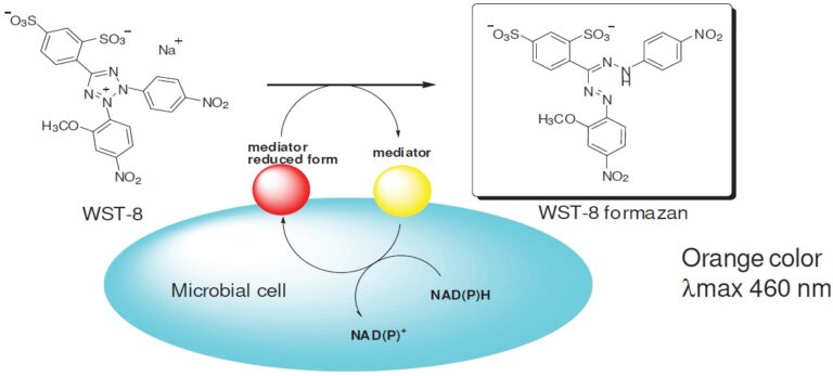
Fig. 1 Bacterial cell viability detection mechanism.

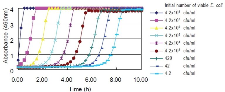
Fig. 2 Correlation between initial number of E. coli and time-dependent O.D. increase. The initial number of viable E.coli were determined by a colony counting method.

Fig. 3 Correlation between the initial number of SA and time-dependent O.D. increase. The initial number of viable SA were determined by the colony counting method.

Fig. 4 Influence of culture media or substances used for bacterial cell culture.
The data indicated that WST is less sensitive to various culture media or substances which are used for bacterial cell culture. WST is a better tetrazolium salt than XTT for bacterial cell viability assays.
Technical info

Table 6 Initial cell number can reach O.D. 0.5 with 1-hour and 4-hour incubation.
The initial cell number of each microorganism was determined by colony counting. Each microorganism cell culture was diluted with medium and 190 μl of the cell culture was added to each well. Then 10 μl of assay solution was added. The cells were incubated at 30oC or 37oC for 1 hour and 4 hours to determine how many cells are required to reach O.D.=0.5 at 460 nm.
Application
General Procedure 2
Determination of the susceptibility of Staphylococcus aureus to oxacillin
Oxacillin: antimicrobial agent: 0-64 μg/ml
Microorganism: Staphylococcus aureus (SA)
Methicillin-resistant Staphylococcus aureus (MRSA)
1. Culture SA or MRSA with Mueller-Hintonmedium containing various concentrations of Oxacillin for 6 hours at 35ºC.
2. Add Microbial Viability Assay solution equal to 1/20 the volume of the culture medium.
3. Incubate for 2 hours at 35ºC.
4. Measure the O.D. at 450 nm to determine the MIC (Minimum inhibitory concentration).

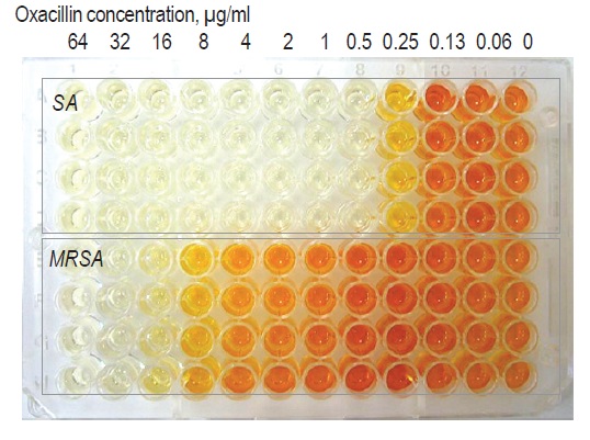
Fig. 4 Susceptibility test of SA and MRSA against Oxacillin*.
The data indicated that MRSA has lower susceptibility than SA. The MICs of MRSA (32 μg/ml) and SA (0.5 μg/ml) are close to the MICs determined by the CLSI (Clinical and Laboratory Standards Institute) method.
References
2. T. Tsukatani, et al., Colorimetric microbial viability assay based on reduction of water-soluble tetrazolium salts for antimicrobial susceptibility testing and screening of antimicrobial substances. Anal Biochem. 2009;393:117-125.
3. T. Tsukatani, et al., Determination of water-soluble vitamins using a colorimetric microbial viability assay based on the reduction of water-soluble tetrazolium salts. Food Chem.2011;127:711-715
4. T. Tsukatani, et al., Comparison of the WST-8 colorimetric method and the CLSI broth microdilution method for susceptibility testing against drug-resistant bacteria. J Microbiol Methods. 2012;90:160-166
5. Jeffrey C. Pommerville. 2014. Fundamentals of Microbiology: Body Systems Edition (3rd Third Edition), p.339. Burlington, MA: Jones & Bartlett Learning.