Biology reagent for cells, Tissuestaining agent, for Cell staining
Biology reagent for cells, Tissuestaining agent, for Cell staining
Reagent for cell biology Fluorescent stain
M2392 / Methylene Blue Solution [for Cell Staining] 100mL [M2392]
A3396 / Acridine Orange Solution [for Cell Staining] 5 mL
Acridine orange is a nucleic acid stain used to stain dead cells. It is taken up at a ratio of 1 to 3 base pairs of double-stranded DNA and emits green fluorescence (Ex: 500 nm, Em: 520 nm). When bound to RNA or single-stranded DNA, it emits red fluorescence (Ex: 460 nm, Em: 650 nm). Since acridine orange is mutagenic, we offer it as a solution type that can prevent droplets during weighing.
O0483 / Oil Red O [for Biochemical Research] 25g
Oil Red O is one of the azo pigments widely used for the detection of adipocytes and triglycerides for a long time, and stains low-polarity lipids in red. It is easy to use, it can be stained simply by adding a stain solution and then washed, and it is easy to visually identify lipids. Furthermore, it is also possible to quantify the amount of lipid accumulation by eluting the dye with isopropanol after staining and measuring the absorbance.
Product information
Biology reagent for cells, Tissuestaining agent, for Cell staining
Cell Staining Dye
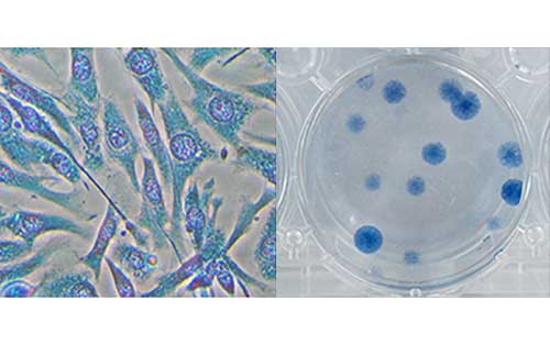
Methylene Blue Solution (Methanol Solution) [M2392] fixes and stains cells at the same time, so you can save fixation before staining. It stains nuclei blue.
Product
M2392 Methylene Blue Solution (Methanol Solution) [for Cell Staining]
Application
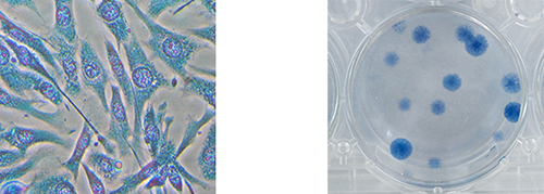
Figure. NIH/3T3 cells stained by the above method
1. Culture cells in a 6-well plate.
2. Remove medium from the plate and wash it with PBS(-) twice.
3. Remove PBS(-) from it, add 1mL of M2392 and stain cells for 15 minutes.
4. Remove M2392 from it and wash it with deionized water twice.
Please adjust staining time and volume according to cells.
Because some cells need to be fixed separately, preliminary tests should be performed.
Dead Cell Staining Dye
Acridine orange is a nucleic acid staining dye that is used to identify dead cells. It intercalates with the double-stranded DNA base pairs at a ratio of 1:3 and is capable of emitting green fluorescence (Ex: 500 nm, Em: 520 nm). It also emits red fluorescence (Ex: 460 nm, Em: 650 nm) when bound to RNA or single-stranded DNA. Since acridine orange is mutagenic, this product is supplied as a solution that prevents it from splashing during weighing.
Product
A3396 Acridine Orange Solution [for Cell Staining]
Application: The method of staining cells by A3396

NIH3T3 cells stained by A3396
1. Remove the medium from the culture plate and wash the cells twice with cold PBS(-). Remove the PBS(-).
2. Add PBS(-) and Acridine Orange solution [A3396] (1/50th of the volume of the added PBS(-)) and incubate for 15 minutes.
3. Remove the staining solution and wash the cells twice with PBS(-).
4. Add PBS(-) and observe the cells under a fluorescence microscope.
Please adjust the staining duration and the volume of the solution according to the cell density.
Some cells may require prior fixation; therefor optimization of the protocol according to your need is recommended.
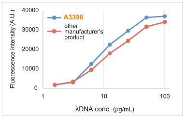
λDNA was stained using either the acridine orange solution [A3396] or the other manufacturer's product, and the fluorescence measured from two experiments was compared (Ex:500nm, Em:520nm).
The results indicated that A3396 stained better than the other manufacturer's product.
Cellular Lipid Staining Dye
Oil red O is a diazo dye which has been widely used for staining fat cell and lipids since long ago. This lysochrome (fat-soluble dye) red dye can be used for staining of neutral triglycerides and low polar lipids. Oil Red O staining is done on fresh or frozen samples, since alcohol fixation removes lipid. Its usage is simple, and requires only washing after adding the staining solution, which makes it easy to identify lipids visually.
Furthermore, accumulation of lipids can be quan-tified by eluting the dye using isopropanol after staining and measuring the absorbance.
Product
O0483 Oil Red O [for Biochemical Research]
Application
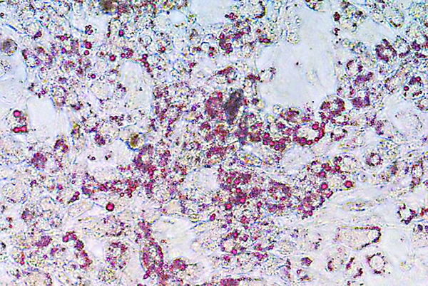
10 days later of addition of media for differentiation to 3T3-L1 cell and then stained by 1mg/mL of O0483.
%20--%3e%3csvg%20version='1.1'%20id='レイヤー_1'%20xmlns='http://www.w3.org/2000/svg'%20xmlns:xlink='http://www.w3.org/1999/xlink'%20x='0px'%20y='0px'%20viewBox='0%200%2055%2063.3'%20style='enable-background:new%200%200%2055%2063.3;'%20xml:space='preserve'%3e%3cstyle%20type='text/css'%3e%20.st0{fill:none;}%20.st1{fill:none;stroke:%23FFFFFF;stroke-miterlimit:10;}%20.st2{fill:%23EAADC6;}%20%3c/style%3e%3cg%20id='レイヤー_2_00000074435297100839857730000016701895290514446243_'%3e%3cg%20id='レイヤー_1-2'%3e%3crect%20class='st0'%20width='55'%20height='63.3'/%3e%3cg%20id='Layer_1'%3e%3cg%3e%3cpath%20class='st1'%20d='M16.3,6.3h22.5c1.6,0,3,1.3,3,3v50.1c0,1.6-1.3,3-3,3H16.3c-1.6,0-3-1.3-3-3V9.2%20C13.3,7.6,14.6,6.3,16.3,6.3z'/%3e%3cpath%20class='st1'%20d='M21.9,1h11.3c0.6,0,1.1,0.5,1.1,1.1v4.2H20.8V2.1C20.8,1.5,21.3,1,21.9,1z'/%3e%3crect%20x='16.2'%20y='9.9'%20class='st2'%20width='22.2'%20height='7.7'/%3e%3crect%20x='16.2'%20y='51.7'%20class='st2'%20width='22.2'%20height='7.7'/%3e%3cpolygon%20class='st2'%20points='29.9,19.9%2022.5,34.5%2026.9,36.4%2024.8,48.8%2032.5,33.9%2027.8,32.1%20'/%3e%3cg%3e%3cline%20class='st1'%20x1='25.2'%20y1='13.8'%20x2='29.4'%20y2='13.8'/%3e%3cline%20class='st1'%20x1='27.4'%20y1='11.7'%20x2='27.4'%20y2='15.9'/%3e%3c/g%3e%3cline%20class='st1'%20x1='25.2'%20y1='55.5'%20x2='29.4'%20y2='55.5'/%3e%3c/g%3e%3c/g%3e%3c/g%3e%3c/g%3e%3c/svg%3e)
%20rotate(-58.77)'/%3e%3cellipse%20class='cls-2'%20cx='28.07'%20cy='31.88'%20rx='31.38'%20ry='12.11'%20transform='translate(-12.46%2019.17)%20rotate(-31.23)'/%3e%3ccircle%20class='cls-1'%20cx='19.93'%20cy='9.05'%20r='3.48'/%3e%3ccircle%20class='cls-1'%20cx='51.74'%20cy='38.02'%20r='3.48'/%3e%3ccircle%20class='cls-1'%20cx='16.88'%20cy='48.55'%20r='3.48'/%3e%3ccircle%20class='cls-1'%20cx='28.07'%20cy='31.88'%20r='5.14'/%3e%3c/g%3e%3c/g%3e%3c/svg%3e)
