Reagent for cell biology, Fluorescent stain
Reagent for cell biology, Fluorescent stain
G0406 / Goat Anti-Mouse IgG FITC Conjugate (green fluorescence)
G0453 / Goat Anti-Mouse IgM FITC Conjugate (green fluorescence)
G0452 / Goat Anti-Rabbit IgG FITC Conjugate (green fluorescence)
S0966 / Streptavidin FITC Conjugate (green fluorescence)
G0569 / Goat Anti-Mouse IgG R-PE Conjugate (red fluorescence)
G0577 / Goat Anti-Rabbit IgG R-PE Conjugate (red fluorescence)
T3885 / Streptavidin R-PE Conjugate (red fluorescence)
G0505 / Goat Anti-Mouse IgG DTBTA-Eu3 + Conjugate (red fluorescence)
G0506 / Goat Anti-Rabbit IgG DTBTA-Eu3 + Conjugate (red fluorescence)
S0993 / Streptavidin DTBTA-Eu3 + Conjugate (red fluorescence)
A2412 / DAPI · 2HC (blue fluorescence)
H1343 / Bisbenzimide H 33258 Hydrate (blue fluorescence)
| Product code | Product name | Package | SDS / Protocol |
|---|---|---|---|
| G0406 | Goat Anti-Mouse IgG FITC Conjugate (Green Gluorescence) | 1 vial | SDSProtocol |
| G0453 | Goat Anti-Mouse IgM FITC Conjugate (Green Gluorescence) | 1 vial | SDSProtocol |
| G0452 | Goat Anti-Rabbit IgG FITC Conjugate (Green Gluorescence) | 1 vial | SDSProtocol |
| S0966 | Streptavidin FITC Conjugate (Green Gluorescence) | 1 vial | SDSProtocol |
| G0569 | Goat Anti-Mouse IgG R-PE Conjugate (Red Gluorescence) | 1 vial | SDSProtocol |
| G0577 | Goat Anti-Rabbit IgG R-PE Conjugate (Red Gluorescence) | 1 vial | SDSProtocol |
| T3885 | Streptavidin R-PE Conjugate (Red Gluorescence) | 1 vial | SDSProtocol |
| G0505 | Goat Anti-Mouse IgG DTBTA-Eu3+ Conjugate (Red Gluorescence) | 1 vial | SDSProtocol |
| G0506 | Goat Anti-Rabbit IgG DTBTA-Eu3+ Conjugate (Red Gluorescence) | 1 vial | SDSProtocol |
| S0993 | Streptavidin DTBTA-Eu3+ Conjugate (Red Gluorescence) | 1 vial | SDSProtocol |
| A2412 | DAPI·2HC (Blue Gluorescence) | 5mg | SDSProtocol |
| H1343 | Bisbenzimide H 33258 Hydrate (Blue Gluorescence) | 25mg | SDSProtocol |
Product information
Reagent for cell biology, Fluorescent stain
Applications
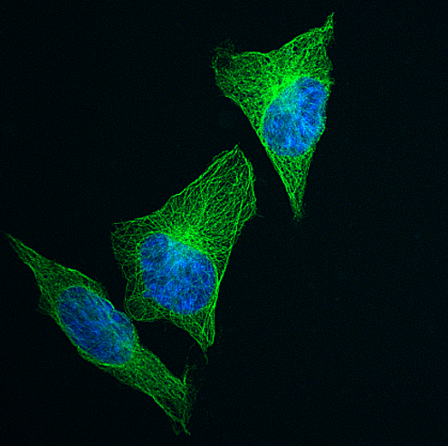
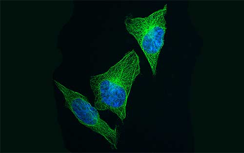
(A) The HeLa cells were incubated with properly diluted primary antibody (Mouse Anti α-Tubulin IgG) and were further incubated with Goat Anti-Mouse IgG Biotin Conjugate [G0387] and Streptavidin FITC Conjugate [S0966] ( green fluorescence ). And then the nuclei was stained with DAPI·2HCl [A2412] ( blue fluorescence ) .
(Laser Scanning Microscope: Olympus FLUOVIEW FV 3000)
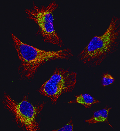
(B) The nuclei of HeLa cells was stained with Bisbenzimide H 33258 [H1343] ( blue fluorescence ). α-Tubulin was stained with anti-α-tubulin antibody and Goat Anti-Mouse IgG Biotin Conjugate [G0387] and Streptavidin R-PE Conjugate [T3885] ( red fluorescence ). Mitochondria was stained with primary antibody and Goat Anti-Rabbit IgG FITC Conjugate [G0452] ( green fluorescence )**
(Laser Scanning Microscope: Olympus FLUOVIEW FV 3000)
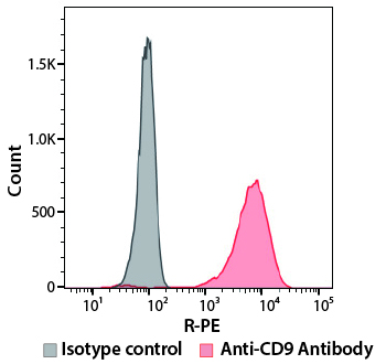
(C) The HeLa cells were incubated with Mouse Anti-CD9 Antibody (red line) or Mouse IgG2aκ isotype control (black line). Subsequently, both were stained with Goat Anti-Mouse IgG Biotin Conjugate [G0387] and Streptavidin R-PE Conjugate [T3885].
(Flow cytometer: Sysmex RF-500)
**Please refer to the product page for staining procedure.
R-PE/FITC-labeled anti-Mouse IgG or anti-Rabbit IgG antibodies and streptavidins can be used for fluorescence immunostaining and flow cytometry.
%20--%3e%3csvg%20version='1.1'%20id='レイヤー_1'%20xmlns='http://www.w3.org/2000/svg'%20xmlns:xlink='http://www.w3.org/1999/xlink'%20x='0px'%20y='0px'%20viewBox='0%200%2055%2063.3'%20style='enable-background:new%200%200%2055%2063.3;'%20xml:space='preserve'%3e%3cstyle%20type='text/css'%3e%20.st0{fill:none;}%20.st1{fill:none;stroke:%23FFFFFF;stroke-miterlimit:10;}%20.st2{fill:%23EAADC6;}%20%3c/style%3e%3cg%20id='レイヤー_2_00000074435297100839857730000016701895290514446243_'%3e%3cg%20id='レイヤー_1-2'%3e%3crect%20class='st0'%20width='55'%20height='63.3'/%3e%3cg%20id='Layer_1'%3e%3cg%3e%3cpath%20class='st1'%20d='M16.3,6.3h22.5c1.6,0,3,1.3,3,3v50.1c0,1.6-1.3,3-3,3H16.3c-1.6,0-3-1.3-3-3V9.2%20C13.3,7.6,14.6,6.3,16.3,6.3z'/%3e%3cpath%20class='st1'%20d='M21.9,1h11.3c0.6,0,1.1,0.5,1.1,1.1v4.2H20.8V2.1C20.8,1.5,21.3,1,21.9,1z'/%3e%3crect%20x='16.2'%20y='9.9'%20class='st2'%20width='22.2'%20height='7.7'/%3e%3crect%20x='16.2'%20y='51.7'%20class='st2'%20width='22.2'%20height='7.7'/%3e%3cpolygon%20class='st2'%20points='29.9,19.9%2022.5,34.5%2026.9,36.4%2024.8,48.8%2032.5,33.9%2027.8,32.1%20'/%3e%3cg%3e%3cline%20class='st1'%20x1='25.2'%20y1='13.8'%20x2='29.4'%20y2='13.8'/%3e%3cline%20class='st1'%20x1='27.4'%20y1='11.7'%20x2='27.4'%20y2='15.9'/%3e%3c/g%3e%3cline%20class='st1'%20x1='25.2'%20y1='55.5'%20x2='29.4'%20y2='55.5'/%3e%3c/g%3e%3c/g%3e%3c/g%3e%3c/g%3e%3c/svg%3e)
%20rotate(-58.77)'/%3e%3cellipse%20class='cls-2'%20cx='28.07'%20cy='31.88'%20rx='31.38'%20ry='12.11'%20transform='translate(-12.46%2019.17)%20rotate(-31.23)'/%3e%3ccircle%20class='cls-1'%20cx='19.93'%20cy='9.05'%20r='3.48'/%3e%3ccircle%20class='cls-1'%20cx='51.74'%20cy='38.02'%20r='3.48'/%3e%3ccircle%20class='cls-1'%20cx='16.88'%20cy='48.55'%20r='3.48'/%3e%3ccircle%20class='cls-1'%20cx='28.07'%20cy='31.88'%20r='5.14'/%3e%3c/g%3e%3c/g%3e%3c/svg%3e)
