CUBIC Series

CUBIC Series
CUBIC is a tissue transparency technology developed by Professor Yasumi Ueda and his colleagues at the RIKEN Center for Bioinformatics and the University of Tokyo Graduate School of Medicine, which enables comprehensive analysis of cellular functions at single-cell resolution through transparency of the whole brain and body of mice. We provide CUBIC reagents for use in this tissue transparency.
| Product code | Product name | Package | SDS / Protocol |
|---|---|---|---|
| T3740 | Tissue-Cleaning Reagent CUBIC-L(for delipidation and decoloring) | 25 ml / 100 ml / 500 ml | SDSProtocol |
| T3983 | Tissue-Clearing Reagent CUBIC-R+(N) [for RI matching] | 25 ml / 100 ml / 500 ml | SDSProtocol |
| T3741 | Tissue-Clearing Reagent CUBIC-R+(M) [for RI matching] | 25 ml / 100 ml | SDSProtocol |
| T3780 | Tissue-Clearing Reagent CUBIC-B [for decalcification] | 25 ml / 100 ml | SDSProtocol |
| T3781 | Tissue-Clearing Reagent CUBIC-HL [for highly fatty tissue and quenching autofluorescence] | 25 ml / 100 ml | SDSProtocol |
| T3782 | Tissue-Clearing Reagent CUBIC-P [efficiently aids perfusion fixation] | 25 ml / 100 ml | SDSProtocol |
| T3866 | Tissue-Clearing Reagent CUBIC-X1 [for tissue expansion] | 25 ml / 100 ml | SDSProtocol |
| T3867 | Tissue-Clearing Reagent CUBIC-X2 [for RI matching while keeping the expanded size] | 25 ml / 100 ml | SDSProtocol |
| M3294 | Mounting Solution (RI 1.520) [for CUBIC-R+] | 50 ml | SDSProtocol |
| M3292 | Mounting Solution (RI 1.467) [for CUBIC-X2] | 50 ml | SDSProtocol |
Product information
Animal tissue clearing reagent CUBIC Series
• BASIC PROTOCOL: Soaking in only two products makes mouse whole-body or animal tissues clear. CUBIC-L (for delipidation and decoloring) and CUBIC-R+ (for RI matching).
CUBIC-L: for delipidation and decoloring
CUBIC-R+: for RI matching
• OPTIONAL PROTOCOL: The following products can easily clear tissues which were previously difficult to clear.
CUBIC-B: for bone
CUBIC-HL: for highly fatty tissues and quenching autofluorescence
CUBIC-P: for mouse perfusion efficiently aids with perfusion fixation
• EXPANSION PROTOCOL: The following products can clear tissues with expansion.
CUBIC-X1: for expansion tissues
CUBIC-X2: for RI matching with keeping the expanded size
• Preserve the fluorescent protein signals except CUBIC-HL.
• Shorter sample treatment period.
• Using light-sheet fluorescent microscopy (LSFM) or confocal laser-scanning microscopy (CLSM) enables the whole-organ/body imaging at a cellular resolution.
Example
Mouse whole-body clearing
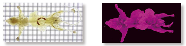
Figure 1. Whole-body clearing (Left), Whole-body clearing with propidium iodide staining (Right)
Mouse whole-body clearing procedure
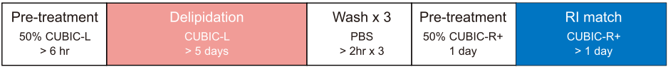
*If you want, please do staining.
Mouse whole-organ clearing
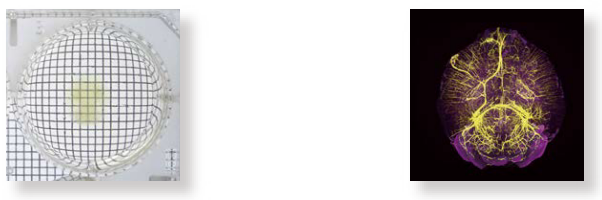
Figure 2. Whole-brain clearing (Left),
Whole-brain clearing with RedDot 2 staining and immunostaining (Right)
Mouse whole-organ clearing procedure
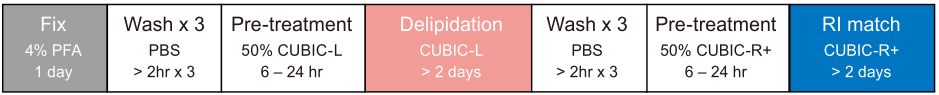
*If you want, please do staining.
The efficient clearing of adult mouse (more than 6-week-old) whole-body or organ samples
The efficient clearing procedure of adult mouse (more than 6-week-old) whole-body or organ samples

*If you want, please do staining.
The clearing of mouse body or tissues including bone
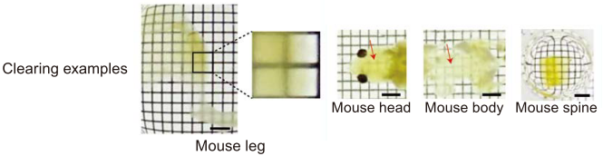
The clearing procedure of mouse body or tissues including bone

The clearing of human brain tissue large blocks
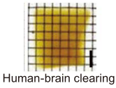
The clearing procedure of human brain tissue large blocks

The clearing of aggressive human tissue
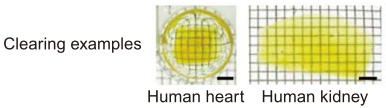
The clearing procedure of aggressive human tissue

How to use CUBIC reagents
References
Mouse Whole Body, Brain, Lung, Liver, Leg, Kidney, Marmoset Brain, Human Brain, Kidney, Liver, Lung Clearing
[Immunohistochemistry after CUBIC protocol]
Chemical Landscape for Tissue Clearing based on Hydrophilic Reagents
K. Tainaka, T. C. Murakami, E. A. Susaki, C. Shimizu, R. Saito, K. Takahashi, A. Hayashi-Takagi, H. Sekiya, Y. Arima, S. Nojima, M. Ikemura, T. Ushiku, Y. Shimizu, M. Murakami, K. F. Tanaka, M. Iino, H. Kasai, T. Sasaoka, K. Kobayashi, K. Miyazono, E. Morii, T. Isa, M. Fukayama, A. Kakita, H. R. Ueda, Cell Reports 2018, 24, 2196.
Mouse Whole Body, Brain, Lung Clearing
Whole-Body Profiling of Cancer Metastasis with Single-Cell Resolution
S. I. Kubota, K. Takahashi, J. Nishida, Y. Morishita, S. Ehata, K. Tainaka, K. Miyazono, H. R. Ueda, Cell Reports 2017, 20, 236.
Mouse Brain, Marmoset Brain Clearing
Whole-Brain Imaging with Single-Cell Resolution Using Chemical Cocktails and Computational Analysis
E. A. Susaki, K. Tainaka, D. Perrin, F. Kishino, T. Tawara, T. M. Watanabe, C. Yokoyama, H. Onoe, M. Eguchi, S. Yamaguchi, T. Abe, H. Kiyonari, Y. Shimizu, A. Miyawaki, H. Yokota, H. R. Ueda, Cell 2014, 157, 726.
With CUBIC Perfusion,
Mouse Whole Body, Heart, Lung, Kidney, Liver Clearing
Whole-Body Imaging with Single-Cell Resolution by Tissue Decolorization
K. Tainaka, S. I. Kubota, T. Q. Suyama, E. A. Susaki, D. Perrin, M. Ukai-Tadenuma, H. Ukai, H. R. Ueda, Cell 2014, 159, 911.
Using CUBIC-X1 and CUBIC-X2,
Mouse Brain Expansion
A three-dimensional single-cell-resolution whole-brain atlas using CUBIC-X expansion microscopy and tissue clearing
T. C. Murakami, T. Mano, S. Saikawa, S. A. Horiguchi, D. Shigeta, K. Baba, H. Sekiya, Y. Shimizu, K. F. Tanaka, H. Kiyonari, M. Iino, H. Mochizuki, K. Tainaka, H. R. Ueda, Nat. Neurosci. 2018, 21, 625.
Application to Pathological Tissue Diagnosis
CUBIC pathology: three-dimensional imaging for pathological diagnosis
S. Nojima, E. A. Susaki, K. Yoshida, H. Takemoto, N. Tsujimura, S. Iijima, K. Takachi, Y. Nakahara, S. Tahara, K. Ohshima, M. Kurashige, Y. Hori, N. Wada, J. Ikeda, A. Kumanogoh, E. Morii, H. R. Ueda, Sci. Rep. 2017, 7, 9269.
%20--%3e%3csvg%20version='1.1'%20id='レイヤー_1'%20xmlns='http://www.w3.org/2000/svg'%20xmlns:xlink='http://www.w3.org/1999/xlink'%20x='0px'%20y='0px'%20viewBox='0%200%2055%2063.3'%20style='enable-background:new%200%200%2055%2063.3;'%20xml:space='preserve'%3e%3cstyle%20type='text/css'%3e%20.st0{fill:none;}%20.st1{fill:none;stroke:%23FFFFFF;stroke-miterlimit:10;}%20.st2{fill:%23EAADC6;}%20%3c/style%3e%3cg%20id='レイヤー_2_00000074435297100839857730000016701895290514446243_'%3e%3cg%20id='レイヤー_1-2'%3e%3crect%20class='st0'%20width='55'%20height='63.3'/%3e%3cg%20id='Layer_1'%3e%3cg%3e%3cpath%20class='st1'%20d='M16.3,6.3h22.5c1.6,0,3,1.3,3,3v50.1c0,1.6-1.3,3-3,3H16.3c-1.6,0-3-1.3-3-3V9.2%20C13.3,7.6,14.6,6.3,16.3,6.3z'/%3e%3cpath%20class='st1'%20d='M21.9,1h11.3c0.6,0,1.1,0.5,1.1,1.1v4.2H20.8V2.1C20.8,1.5,21.3,1,21.9,1z'/%3e%3crect%20x='16.2'%20y='9.9'%20class='st2'%20width='22.2'%20height='7.7'/%3e%3crect%20x='16.2'%20y='51.7'%20class='st2'%20width='22.2'%20height='7.7'/%3e%3cpolygon%20class='st2'%20points='29.9,19.9%2022.5,34.5%2026.9,36.4%2024.8,48.8%2032.5,33.9%2027.8,32.1%20'/%3e%3cg%3e%3cline%20class='st1'%20x1='25.2'%20y1='13.8'%20x2='29.4'%20y2='13.8'/%3e%3cline%20class='st1'%20x1='27.4'%20y1='11.7'%20x2='27.4'%20y2='15.9'/%3e%3c/g%3e%3cline%20class='st1'%20x1='25.2'%20y1='55.5'%20x2='29.4'%20y2='55.5'/%3e%3c/g%3e%3c/g%3e%3c/g%3e%3c/g%3e%3c/svg%3e)
%20rotate(-58.77)'/%3e%3cellipse%20class='cls-2'%20cx='28.07'%20cy='31.88'%20rx='31.38'%20ry='12.11'%20transform='translate(-12.46%2019.17)%20rotate(-31.23)'/%3e%3ccircle%20class='cls-1'%20cx='19.93'%20cy='9.05'%20r='3.48'/%3e%3ccircle%20class='cls-1'%20cx='51.74'%20cy='38.02'%20r='3.48'/%3e%3ccircle%20class='cls-1'%20cx='16.88'%20cy='48.55'%20r='3.48'/%3e%3ccircle%20class='cls-1'%20cx='28.07'%20cy='31.88'%20r='5.14'/%3e%3c/g%3e%3c/g%3e%3c/svg%3e)
